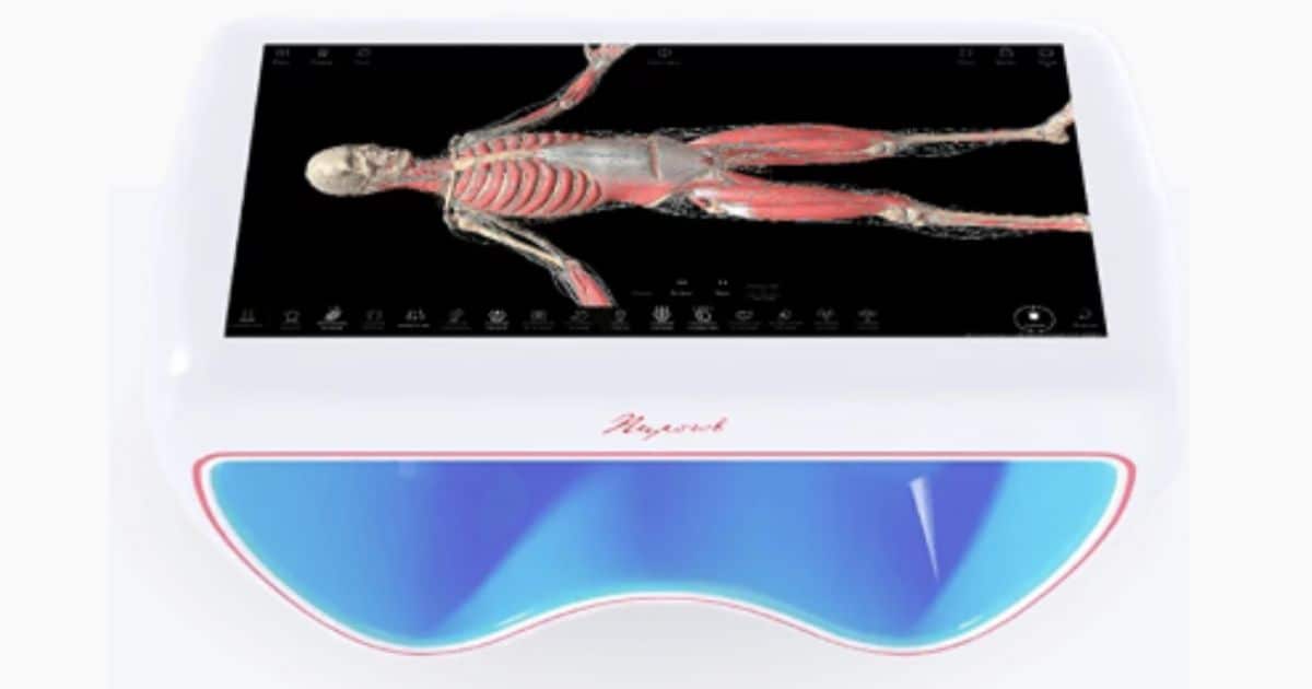
A Virtual Anatomy Table is an innovative medical device designed for the study of human anatomy. It typically consists of a large touchscreen display, sometimes in conjunction with special virtual reality (VR) goggles, allowing medical students, professionals, and researchers to explore internal structures of the human body without the need for real anatomical specimens or body cross-sections. These tables often provide interactive 3D models that users can manipulate, zoom in on, and rotate to examine different anatomical features and systems in detail. They are valuable tools for medical education, surgical planning, and research purposes.
Why are anatomical tables popular in 2024?
Virtual anatomy tables have become popular in recent times due to several reasons:
Technological Advancements: With advancements in virtual and augmented reality technologies, it has become possible to create more realistic and interactive virtual models of human anatomy. This enhances the learning experience and provides a more immersive way to study anatomy compared to traditional methods.
Accessibility: Virtual anatomy tables offer accessibility to anatomy education without the need for expensive anatomical specimens or cadavers. They can be used in various educational settings, including medical schools, hospitals, and research institutions, making anatomy education more widely available.
Safety and Ethical Considerations: Using virtual anatomy tables eliminates the need for handling real human cadavers, addressing safety concerns and ethical considerations associated with traditional dissection methods. This makes virtual anatomy tables an attractive option for medical education programs.
Interactivity and Customization: Virtual anatomy tables allow users to interact with 3D models of anatomical structures, enabling them to manipulate, zoom in on, and explore different layers of the human body. This interactivity facilitates a deeper understanding of complex anatomical concepts and allows for customization of learning experiences based on individual needs.
Integration with Curriculum: Many medical schools and educational institutions are incorporating virtual anatomy tables into their curriculum to enhance anatomy education. The versatility of these tools allows educators to integrate them into existing teaching methods, lectures, and practical sessions, providing students with a comprehensive learning experience.
Pirogov Interactive Table

The Pirogov Anatomy Table, also known as the Anatomage Table, is an advanced virtual dissection table used for medical education, research, and surgical planning. Named after the renowned Russian surgeon Nikolay Pirogov, it offers a comprehensive and immersive platform for studying human anatomy.
The table features a large touchscreen display with high-resolution 3D models of anatomical structures, including organs, tissues, and systems of the human body. Users can interact with the virtual models, manipulate them in real-time, and explore various anatomical layers with precision.
One of the key features of the Pirogov Anatomy Table is its ability to provide realistic simulations of surgical procedures. Surgeons can use the table to plan surgeries, practice techniques, and visualize complex anatomical relationships before performing procedures on real patients. This can improve surgical outcomes and reduce the risk of complications.
The Pirogov Anatomy Table has gained popularity in medical schools, hospitals, and research institutions worldwide due to its effectiveness in anatomy education, surgical training, and patient care. It offers a safe, ethical, and cost-effective alternative to traditional methods of anatomy teaching and surgical planning, making it an invaluable tool in modern medical practice.
